In a collaboration with EPFL Lausanne, ETH Zurich and the College of Southern California researchers on the Paul Scherrer Institute PSI have used X-rays to look inside a microchip with greater precision than ever earlier than. The picture decision of 4 nanometers marks a brand new world file. The high-resolution three-dimensional pictures of the sort they produced will allow advances in each data expertise and the life sciences.
The researchers are reporting their findings within the present subject of the journal Nature.
Since 2010, the scientists on the Laboratory of Macromolecules and Bioimaging at PSI have been creating microscopy strategies with the purpose of manufacturing three-dimensional pictures within the nanometer vary. Of their present analysis, a collaboration with the EPFL and the ETHZ, the Swiss Federal Institutes of Expertise in Lausanne and Zürich, and the College of Southern California, they’ve succeeded for the primary time in taking footage of state-of-the-art laptop chips microchips with a decision of 4 nanometers—a world file.
As a substitute of utilizing lenses, with which pictures on this vary should not at present potential, the scientists resort to a method generally known as ptychography, through which a pc combines many particular person pictures to create a single, high-resolution image. Shorter publicity occasions and an optimized algorithm had been key to considerably bettering upon the world file they themselves set in 2017. For his or her experiments, the researchers used X-rays from the Swiss Mild Supply SLS at PSI.
Between standard X-ray tomography and electron microscopy
Microchips are marvels of expertise. These days, it’s potential to pack greater than 100 million transistors per sq. millimeter into superior built-in circuits—a development that continues to extend. Extremely automated optical programs are used to etch the nanometer-sized circuit traces into silicon blanks in clear rooms.
Layer after layer is added and eliminated till the completed chip, the brains of our smartphones and computer systems, could be lower out and put in. The manufacturing course of is elaborate and complex, and characterizing and mapping the ensuing constructions proves to be simply as troublesome.
Whereas scanning electron microscopes have a decision of some nanometers and are subsequently nicely suited to imaging the tiny transistors and steel interconnects that make up circuits, they’ll solely produce two-dimensional pictures of the floor.
“The electrons don’t travel far enough into the material,” explains Mirko Holler, a physicist at SLS. “To construct three-dimensional images with this technique, the chip has to be examined layer by layer, removing individual layers at the nanometer level—a very complex and delicate process which also destroys the chip.”
Nonetheless, three-dimensional and non-destructive pictures could be produced utilizing X-ray tomography, as a result of X-rays can penetrate supplies a lot additional. This process is much like a CT scan in a hospital. The pattern is rotated and X-rayed from totally different angles. The best way the radiation is absorbed and scattered varies, relying on the inner construction of the pattern. A detector data the sunshine leaving the pattern and an algorithm reconstructs the ultimate 3D picture from it.
“Here we have a problem with the resolution,” explains Holler. “None of the X-ray lenses currently available can focus this radiation in a way that allows such tiny structures to be resolved.”
Ptychography—the digital lens
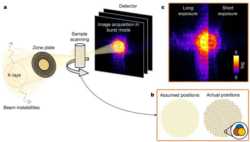
The answer is ptychography. On this approach, the X-ray beam shouldn’t be centered on a nanometer scale; as an alternative, the pattern is moved on a nanometer scale. “Our sample is moved such that the beam follows a precisely defined grid—like a sieve. At each point along the grid, a diffraction pattern is recorded,” explains the physicist.
The space between the person grid factors is lower than the diameter of the beam, so the imaged areas overlap. This produces sufficient data to reconstruct the pattern picture at a excessive decision with the assistance of an algorithm. The reconstruction course of is fairly like utilizing a digital lens.
“Since 2010, we have been steadily perfecting our experimental set-up and the accuracy with which we position our samples. In 2017, we finally succeeded in spatially imaging a computer chip with a resolution of 15 nanometers—a record,” Holler remembers.
Since then, the decision has remained unchanged in our instrument, regardless of additional optimizations within the set-up and the algorithm. “We extended it by one or two nanometers, but that was as far as we could go. Something was limiting us and we had to find out what it was,” he provides.
The seek for the limiting issue
The frilly search lastly started in 2021. Along with Holler and Manuel Guizar-Sicairos, who had each been concerned within the first file, Tomas Aidukas additionally joined the group. The physicist supported the staff along with his programming expertise and developed the brand new algorithm which in the end helped them to attain the breakthrough.
The researchers discovered their first clue after they decreased the publicity time—all of a sudden the diffraction pictures had been sharper. This led them to conclude that the X-ray beam illuminating the pattern was not secure, however as an alternative shifting by tiny quantities—the beam was wobbling.
“This is analogous to photography,” Guizar-Sicairos explains. “When you take a picture at night, you choose a long exposure because of the darkness. If you do this without using a tripod, your movements are transmitted to the camera and the picture will be blurred.”
Alternatively, in the event you select a brief publicity time in order that the sunshine is captured sooner than we transfer, then the picture shall be sharp. “But in that case, the picture might be completely black or noisy, because almost no light can be captured in that short amount of time,” he provides.
The researchers confronted an identical downside. Though their pictures had been now sharp, they contained too little data to reconstruct your entire microchip, due to the quick publicity time.
Shorter publicity time and a brand new algorithm
To resolve the issue, the researchers upgraded their set-up with a sooner detector, additionally developed at PSI. This allowed them to file many pictures at every grid level, every with a brief publicity time.
“A huge mountain of data,” Aidukas provides. When the person pictures are added collectively and superimposed, this leads to the identical blurry picture that was obtained utilizing a protracted publicity time.
“You can think of the X-ray beam as one point on the sample. We now take a whole lot of individual pictures at this particular point,” explains Aidukas. Because the beam is wobbling, every picture will change barely. “In some of the pictures, the beam is in the same position, in others it has moved. We can use these changes to track the actual position of the beam caused by the unknown vibrations.”
The following factor is to cut back the quantity of information. “Our algorithm compares the positions of the beam in the individual images. If the positions are the same, they are put in the same group and added to the sum,” he provides.
By grouping the low-exposure pictures, their data content material could be elevated. In consequence, the researchers are capable of reconstruct a pointy picture with a excessive gentle content material utilizing the flood of short-exposure footage.
The brand new ptychographic approach is a primary method that may also be used at related analysis services. The strategy shouldn’t be confined to microchips, however may also be used on different samples, for instance in supplies science or life sciences.
Extra data:
Tomas Aidukas et al, Excessive-performance 4-nm-resolution X-ray tomography utilizing burst ptychography, Nature (2024). DOI: 10.1038/s41586-024-07615-6
Offered by
Paul Scherrer Institute
Quotation:
New X-ray world file: Wanting inside a microchip with 4 nanometer precision (2024, August 6)
retrieved 6 August 2024
from https://phys.org/information/2024-08-ray-world-microchip-nanometer-precision.html
This doc is topic to copyright. Aside from any truthful dealing for the aim of personal examine or analysis, no
half could also be reproduced with out the written permission. The content material is offered for data functions solely.

